

Advanced Intrauterine Diagnostics & Surgery
Hysteroscopy is a specialized procedure that allows a gynaecologist to look inside the uterus to diagnose and treat causes of abnormal bleeding or fertility issues. Using a thin, lighted tube called a hysteroscope, Dr. Smitha can view the uterine cavity on a high-definition monitor. Because the scope is inserted through the natural opening of the cervix, there are no external incisions or scars.
This procedure serves two purposes: **Diagnostic Hysteroscopy** to identify issues like polyps, fibroids, or structural anomalies, and **Operative Hysteroscopy** to treat those conditions simultaneously. At our clinic, we prioritize patient comfort and precision, using the latest endoscopic technology to provide clear answers and effective surgical solutions with minimal recovery time.
Clinical Applications
- Abnormal Uterine Bleeding
- Removal of Uterine Polyps
- Submucosal Fibroid Removal
- Infertility Evaluation
- Recurrent Miscarriage Investigation
- Removal of Adhesions (Asherman’s)
- Correction of Uterine Septum
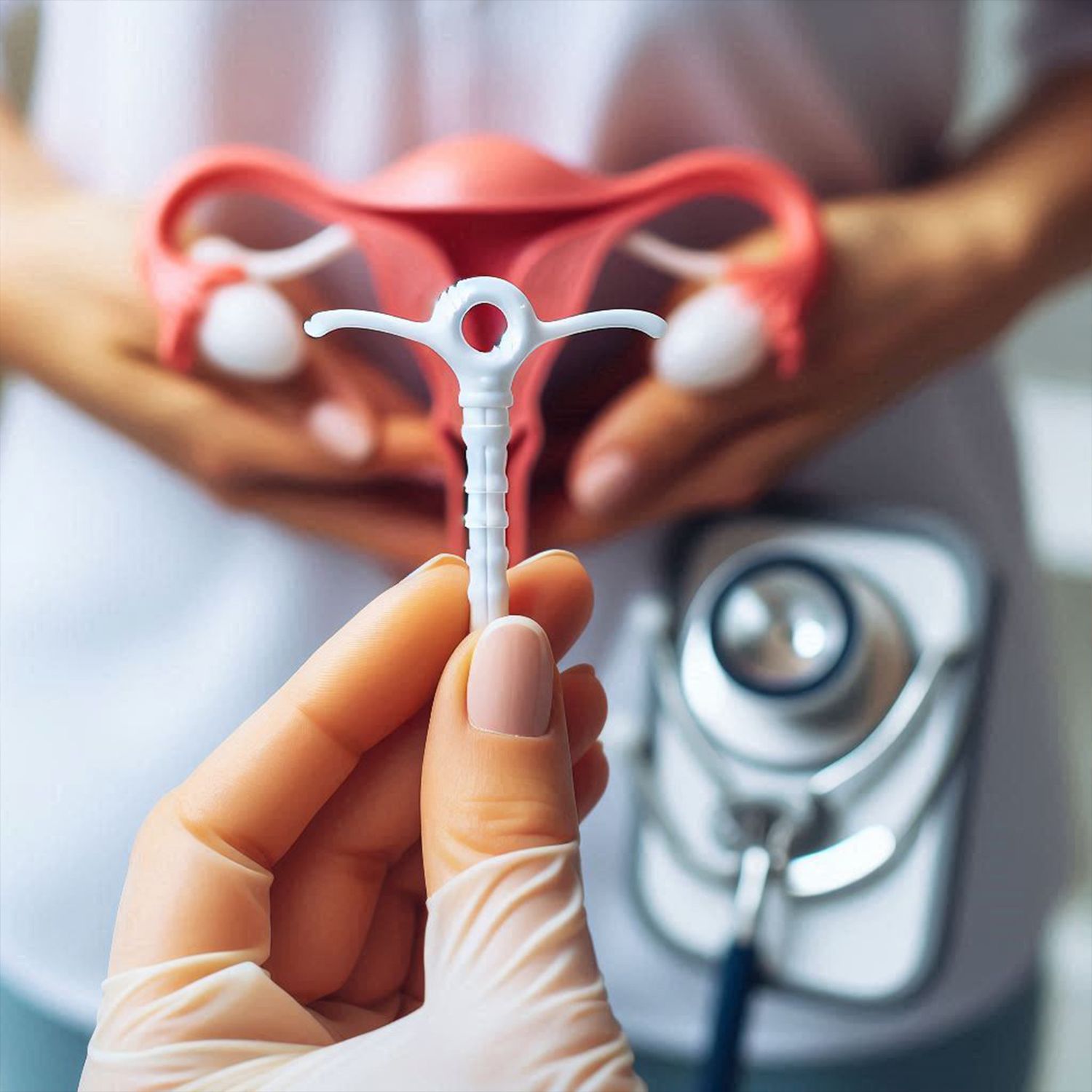
- Displaced IUCD Removal
- Endometrial Biopsy Direction
- Minimal Post-Op Discomfort
- No External Scarring
- Rapid Recovery & Return to Work
The Procedure Protocol
Diagnostic Viewing
The hysteroscope is gently inserted to expand the uterus with saline or gas, providing a clear view of the lining and tubal openings.
Operative Intervention
If an abnormality is found, specialized micro-instruments are passed through the scope to perform the necessary repair immediately.
Post-Op Observation
Most patients spend 1–2 hours in recovery before being discharged the same day with minimal physical restrictions.
Endoscopy Gallery
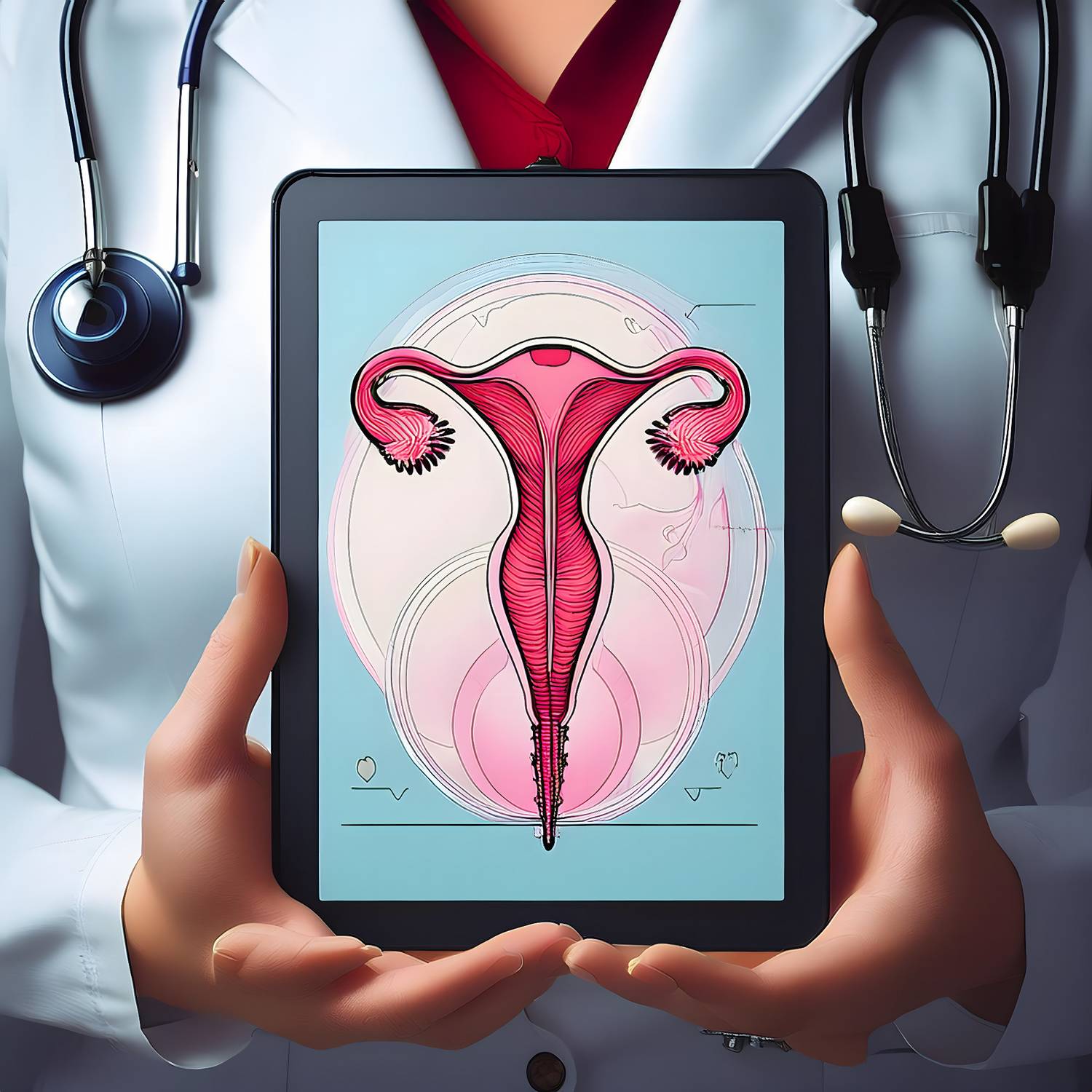
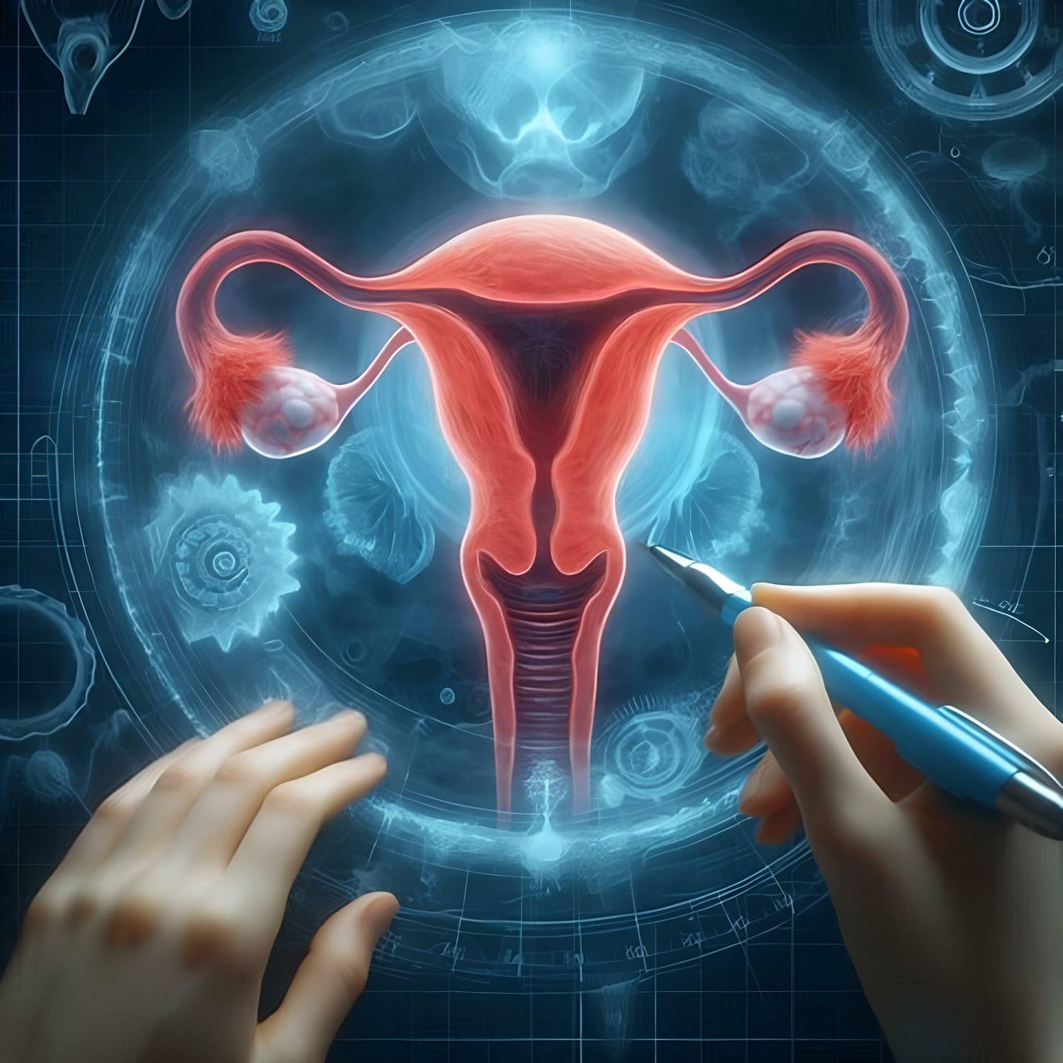
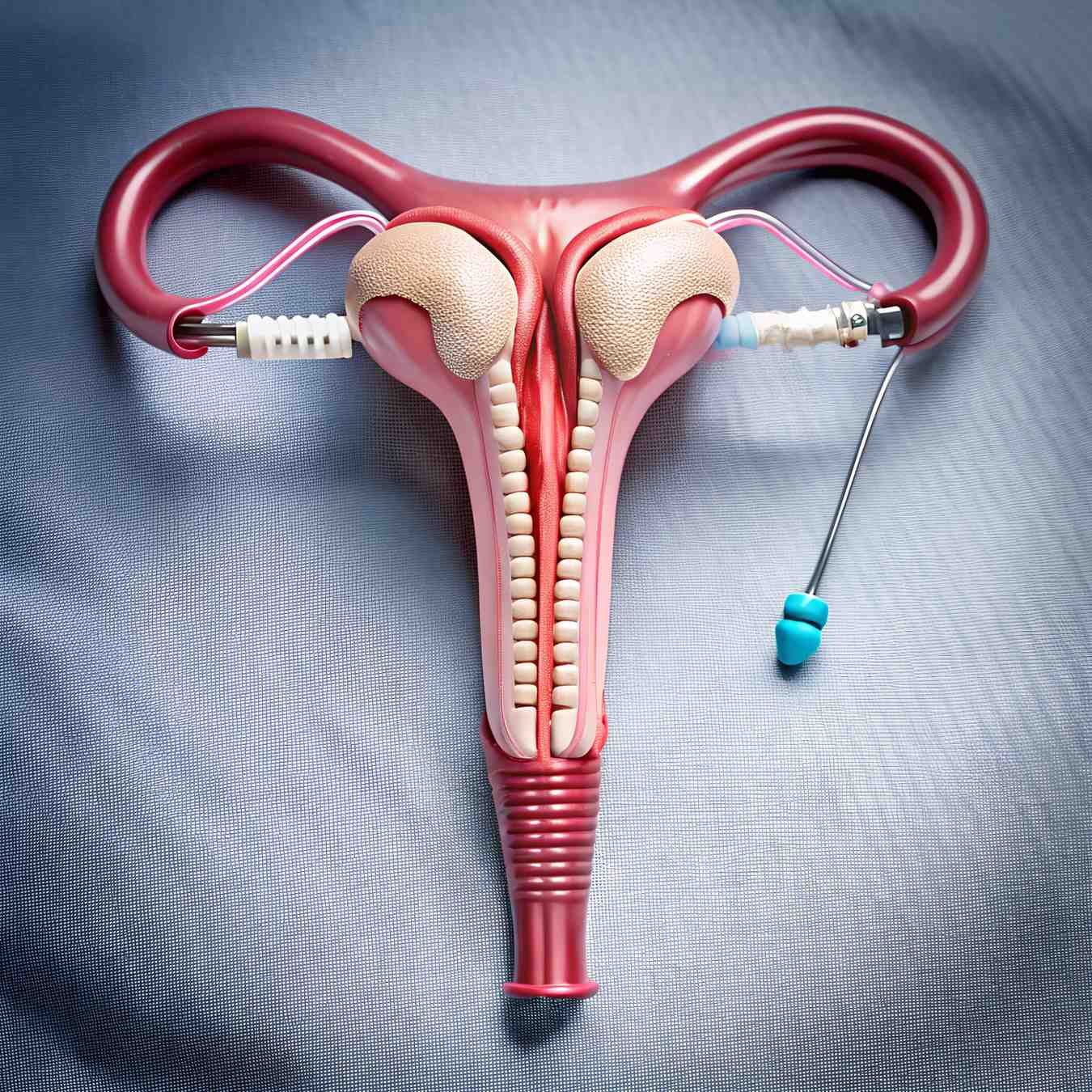
Our Moosapet surgical suite is equipped with advanced Karl Storz endoscopic systems for high-definition intrauterine visualization.

Hysteroscopy FAQ
Common clinical questions regarding the procedure and recovery.
Diagnostic hysteroscopy can often be done with local anesthesia. However, for operative procedures like fibroid removal, we typically use general or spinal anesthesia to ensure complete comfort and safety.
Most patients return to their normal routine the very next day. You may experience some mild cramping or light spotting for 2–3 days, which is entirely normal and manageable with simple medication.
The procedure is usually scheduled for the week following your period. This is when the uterine lining is thinnest, providing the doctor with the clearest possible view of the cavity.